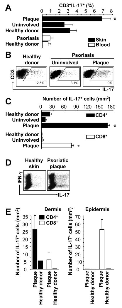
FIGURE 1.
IL-17+ T cells in psoriasis lesions. Skin biopsies and peripheral blood from healthy donors and patients with psoriasis were stained with specific antibodies as described in Materials and Methods. A-E, IL-17+ T cells were analyzed with FACS. A, IL-17+ T cells. Results were expressed as the mean percent of IL-17+ T cells in T cells, error bars indicate SEM, n = 8-10. B, Representative dot-plots are shown. C, Total number of IL-17+ T cells in the skin. Total number of IL-17+ T cells was calculated by multiplying the percentage of IL-17+ T cells by the absolute number of T cells per mm2 of skin, as determined previously (26). Results are expressed as mean number ± SEM, n = 10. D, Coexpression of IL-17 and IFNγ on IL-17+ T cells. Single cell suspensions were made from skin tissues in healthy donors and patients with psoriasis. The expression of IFNγ and IL-17 was analyzed by intracellular cytokine staining gated on CD3+ T cells. E, Total number of IL-17+ T cell subsets in dermis and epidermis. Total number of IL-17+CD4+ and IL-17+CD8+ T cell subsets was calculated by multiplying the percentage of each IL-17+ T cell subset by the absolute number of T cell subsets per mm2 skin (26) in the separated dermis and epidermis. Results are expressed as mean number ± SEM, n = 5. *P < 0.05 (A, C, E) compared to uninvolved and healthy donor skin.

