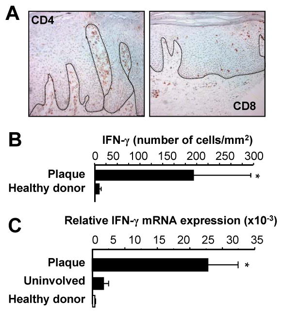
FIGURE 2.

IFNγ+ T cells in psoriasis lesions. A, T cells in psoriasis lesions. Psoriasis plaques were stained with anti-CD4 and anti-CD8. One of 5 representative individuals is shown. B, C, IFNγ+ T cells in psoriasis lesions. IFN-γ+ T cells in psoriatic lesions: T cells (CD45+CD3+) were analyzed by FACS in cell suspensions obtained from healthy skin and psoriatic plaque. B, IFN-γ+ T cells were analyzed by FACS and quantified in the skin. Results were expressed as the mean number per mm2 skin ± SEM. (n = 3). C, Expression of IFN-γ in skin from healthy donors and patients with psoriasis. IFN-γ was quantified by real-time PCR. Results are expressed as the mean values of relative expression ± SEM. (n = 5, *P < 0.03 compared to uninvolved and healthy skin).
