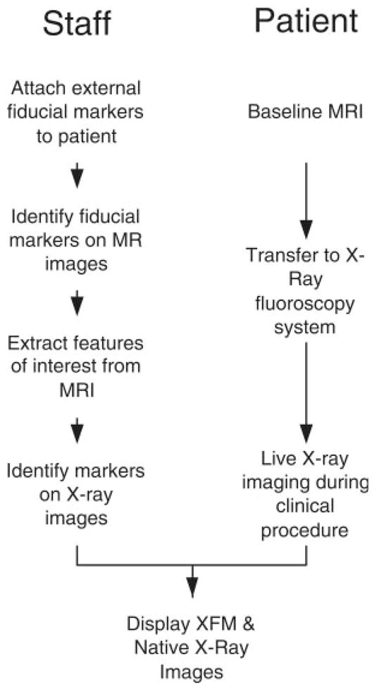
Fig. 1.

Staff and patient workflow during XFM procedures. Baseline MRI is performed after fiducial markers are affixed to patients’ skin. Although the patient is transferred to the X-ray system, the fiducial markers and MRI regions of interest are manually extracted. The fiducial markers are identified on X-ray and image fields are registered. Afterward, the 3D and 2D images are transformed and combined during live X-ray imaging.
