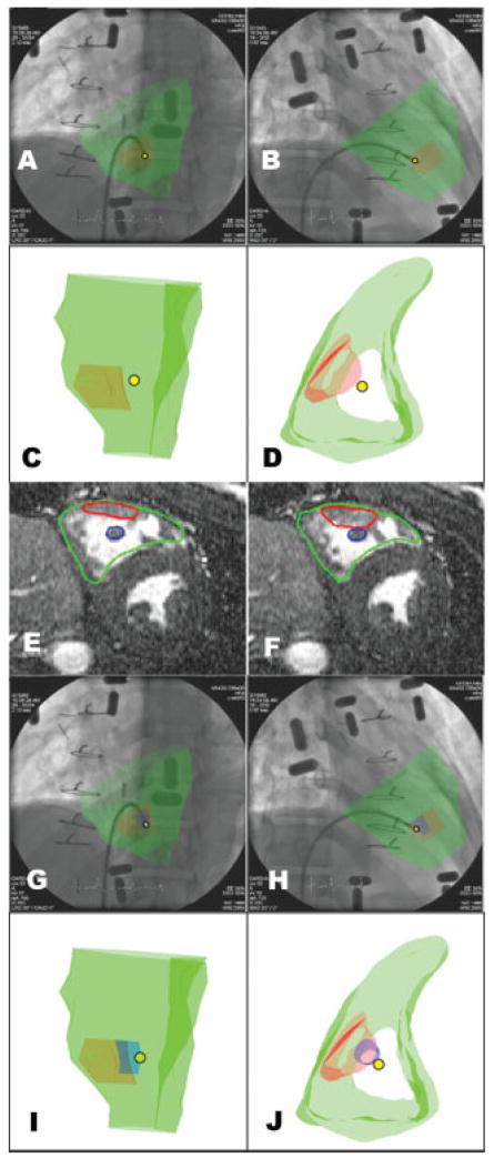
Fig. 3.
Triangulating the position of the bioptome. Panels A–B show two different fluoroscopic projections and corresponding 3D MRI-derived regions of interest (C, D). The right ventricular endocardial border is depicted in green, the mass in red, and the tip of the biopsy forceps is depicted as a yellow dot. The bioptome is discontiguous with the mass (see text). Panels E–J provide an explanation for the spurious normal biopsy finding. Review of MRI (panels E and F) reveals a papillary muscle (blue) posterior to the mass (red). Revised XFM (G–H) incorporates the papillary muscle. Panels I–J shows the 3D image of the mass (red), papillary muscle (blue), and triangulated bioptome position (yellow dot) indicating that the specimens were obtained from the papillary muscle and not from the mass.

