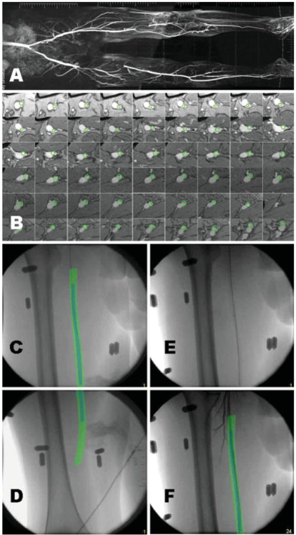
Fig. 6.
XFM-guided recanalization and stenting of R superficial femoral artery occlusion. A: Baseline composite contrast-enhanced MR angiogram shows R SFA occlusion and contra-lateral popliteal and tibial occlusion. B: A descending series of axial T1-weighted MR images permit contrast-independent arterial contours to be segmented (green). C and D: The MRI-derived contours are combined in XFM with the live X-ray images during guidewire recanalization. Note that the automatic gantry position correction permits the same XFM contours to be applied throughout the occlusion, which spans two X-ray fields of view. E: X-ray image after stenting. F: XFM during final poststent angiography.

