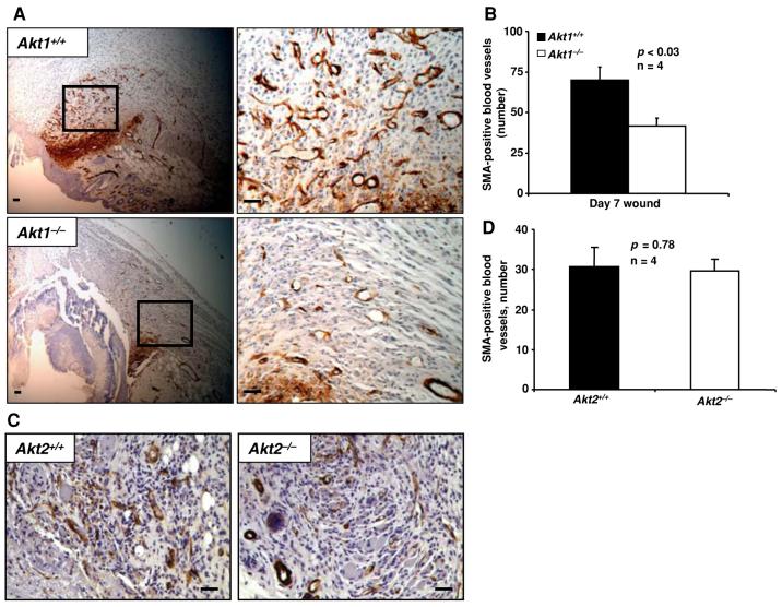
Fig. 4.
Akt1-/- mice, but not Akt2-/-mice, exhibit impaired blood vessel maturation in wounds even on day 7 after wounding. (a) Micrographs of paraffin wound sections from WT and Akt1-/- mice showing SMA-positive blood vessels. (b) SMA-positive blood vessel number as analyzed by manual counting of microscopic fields. (c) Micrographs of paraffin wound sections from WT and Akt2-/- mice showing SMA-positive blood vessels. (d) SMA-positive blood vessel number in WT and Akt2/ wounds as analyzed by manual counting of microscopic fields. Results are expressed as the mean ± sd. Scale bar: 20 μm

