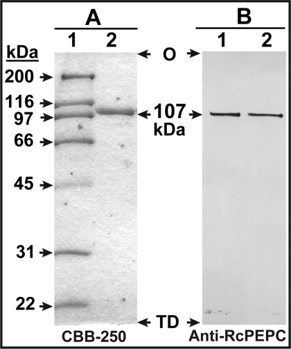
Figure 3. SDS/PAGE and immunoblot analysis of purified PEPC from −Pi Arabidopsis suspension cells.
(A) SDS/PAGE: lane 1 contains various molecular mass standards (3 μg) whereas lane 2 contains 2 μg of the of the pooled peak fractions from the final purification step (Mono Q FPLC). Protein staining was performed using Coomassie Brilliant Blue R-250 (CBB-250). (B) Immunoblotting was performed using an anti-RcPEPC Ab [26]; lanes 1 and 2 contain 25 ng of the final preparation of −Pi Arabidopsis PEPC and homogeneous RcPEPC [26] respectively. O, origin; TD, tracking-dye front.

