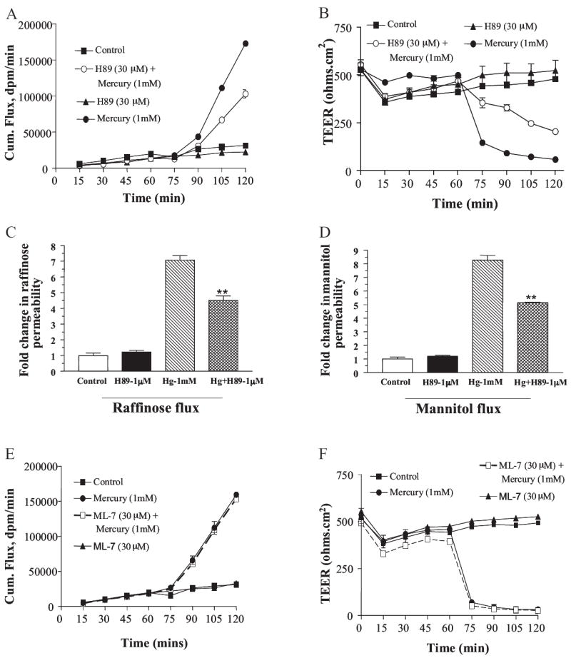
Fig. 2.

Hg2+ activates a signal transduction pathway that involves PKA and is independent of MLCK. Cells were treated with PKA inhibitor H89 and MLCK inhibitor ML-7 at time 0, and mercuric chloride was added after 60 min. A, cumulative mannitol flux as measured at 15-min intervals in presence of 30 μM H89 (n = 6). B, TEER as measured at 15-min intervals in presence of 30 μM H89 (n = 6). C, -fold change in raffinose permeability after addition of Hg2+ and its rescue by H89 at 1 μM concentration (n = 3). D, -fold change in mannitol permeability after addition of Hg2+ and its rescue by H89 at 1 μM concentration (n = 3). Cumulative mannitol flux as measured at 15-min intervals in presence of ML-7 (n = 6). D, TEER as measured at 15-min intervals in presence of ML-7 (n = 6).
