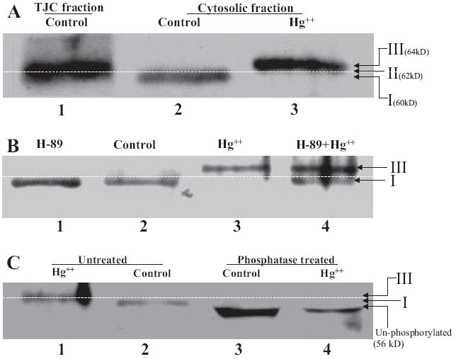
Fig. 6.

Hg2+ triggers phosphorylation and subcellular redistribution of occludin. Western blot analysis of the tight junction and cytosolic fraction of SMG-C6 cells. Cells were treated with PKA inhibitor H89 at time 0, and mercuric chloride was added after 60 min. A, Western blot of the three phosphorylated forms of occludin. Lane 1, form II (62 kDa), found in Triton X-100-insoluble (membrane) fractions of control and Hg2+-treated cells; lane 2, form I (60 kDa), found in Triton X-100-soluble (cytosolic) fraction of control cells; and lane 3, form III (64 kDa), found in Triton X-100-soluble (cytosolic) fractions of cells treated with Hg2+. B, Western blot of occludin in the soluble fraction (Triton X-100-soluble) of SMG-C6 cells. Lane 1, H89 alone; lane 2, control; lane 3, Hg2+; and lane 4, Hg2+ + H89. C, Western blot of occludin in the soluble fraction before phosphatase treatment (lane 1, Hg2+ and lane 2, control) and after phosphatase treatment (lane 3, control and lane 4, Hg2+).
