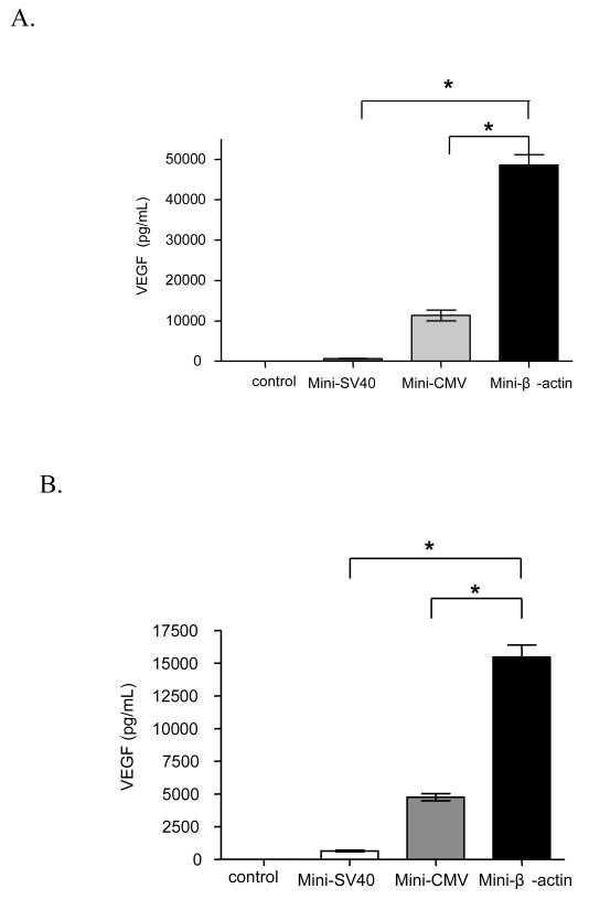
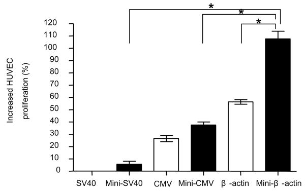
Figure 5.
VEGF protein expression after transfecting C2C12 cells with pMini-SV-VEGF, pMini-CMV-VEGF or pMini-β-VEGF. C2C12 cells without transfection were used as control group. (A) Forty eight hours after transfection. (B) Seventy two hours after transfection. (C) Comparison of VEGF protein expression between minicircle DNA and conventional plasmid under the CMV or chicken β-actin promoter. Data represents mean ± SEM (n = 3). Statistical analysis was done using one-way ANOVA and a Tukey's multiple comparison post-test. * p<0.05.


