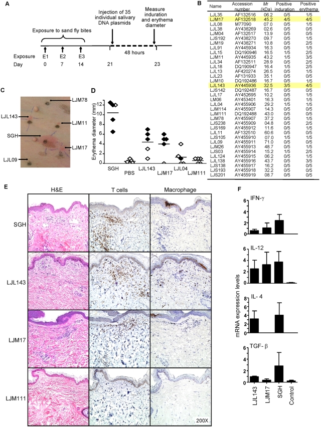
Figure 2. Identification of salivary proteins from Lu. longipalpis that produce a cellular immune response in dogs.
(A) A schematic representation of the reverse antigen screening approach based on the intradermal injection of DNA plasmids in dogs previously exposed to sand fly bites (first exposure, E1; second exposure, E2; third exposure, E3). (B–F) Dogs pre-exposed to sand fly bites were challenged intradermally with DNA plasmids and one pair of salivary gland homogenate (SGH) and PBS (positive and negative controls, respectively) and investigated 48 h post-injection. (B) The number of dogs showing local induration and/or erythema at the site of injection for 35 DNA plasmids coding for secreted salivary molecules. Yellow bars highlight the response of dogs to LJM17 and LJL143. (C) Photograph to demonstrate specificity of the cellular reaction to DNA plasmids and SGH. (D) The diameter of erythema in the absence (◊) or presence (♦) of induration for each dog at the site of injection of SGH, PBS, LJL143 and LJM17 (reactive plasmids) and LJL04 and LJM111 (intermediate and non-reactive plasmids, respectively). (E–F) Skin biopsies (6mm) obtained from injection sites were cut in half and processed for histology and RNA extraction. (E) Representative H&E staining and immunohistochemical labeling of dermal T cells (anti-CD3) and macrophages (Mac387) at the injection sites of SGH, LJL143, LJM17 and LJM111. Note marked dermal infiltrates of inflammatory cells characterized as CD3+ T cells and scattered macrophages (Mac387) in the SGH, LJL143 and LJM17. There is no inflammation with LJM111. (F) Reverse-transcriptase quantitative PCR showing the expression levels of IFN-γ, IL-12, IL-4 and TGF-β for LJL143, LJM17, a pair of SGH and control (a mix of PBS and empty plasmid) 48 h post-injection. Error bars represent means±S.E.

