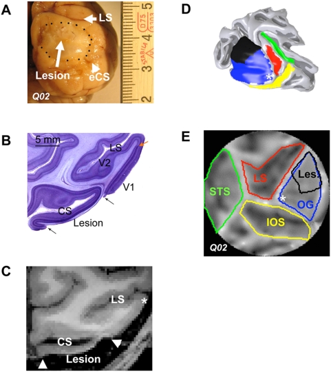
Figure 1. Characterization of the V1 Lesion.
A Picture of Macaque Q02's brain post-mortem. The area of completely denuded gray matter and exposed white matter, is highlighted in the picture by black dots. The dorsal border of the lesion approaches the lunate sulcus (LS) and the ventral border reaches up to the external calcarine (eCS). Major ticks of the scale bar are in centimetres. B Nissl stained axial section (100 µm thick) through the center of Q02's V1 lesioned cortex. Arrows point to the borders of the V1 lesion, which completely destroyed gray matter but largely spared the underlying white matter. Note the characteristic line of Gennari indicating the border between V1 and V2 (red arrow). The V1 cortex surrounding the lesion and the lunate sulcus (LS) containing areas V2 and V3 are not affected by the lesion. C Axial MRI slice through the visual cortex of macaque Q02. The lesioned area is evident in the MR image due to the absence of gray matter. D 3-D reconstruction of the surface of the visual cortex of macaque Q02. The 3D rendering represents the border between gray and white matter. Some of the prominent anatomical landmarks are color coded for easier visualization in the flat map view (see panel E). The V1 lesion shown in black was reconstructed by manually selecting the area devoid of gray matter (panel C). Dorso-ventrally, it starts 1–2 mm ventral to the lunate reaching up to the external calcarine sulcus, and medio-laterally from the edge of the internal calcarine sulcus to ∼14 mm from the intersection of the lunate and inferior occipital sulci (*, fovea). E Flat map of the visual cortex of Macaque Q02. Sulci and gyri are shown as dark and light regions respectively. Sulci and gyri of visual cortex are color-coded in the same way as in the 3-D reconstruction of panel D. Abbreviations: LS: lunate sulcus, eCS: external calcarine sulcus, CS: internal calcarine sulcus, IOS: inferior occipital sulcus, OG: occipital gyrus, STS: superior temporal sulcus Les: Lesion.

