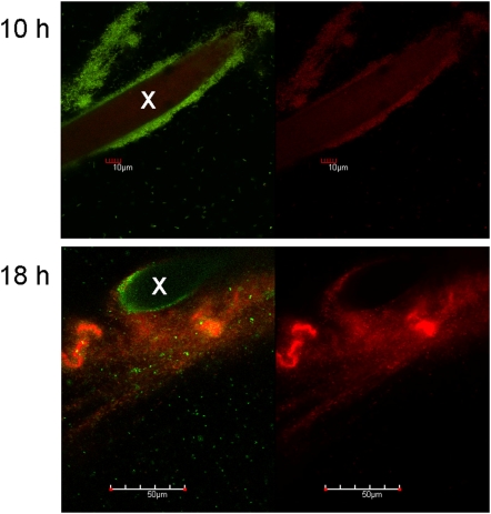
Figure 4. Confocal-microscopical appearance of PE-attached biofilms before and after starvation.
GFP-fluorescent bacteria were grown in presence of a membrane-impermeable DNA stain (BOBO 3) and PE was imaged during the growth (10 h) and during the stationary phase (18 h). The green/red fluorescence image is given (left panel), and solely the red fluorescence channel indicative of dead cells and of extracellular DNA (right panel). Transects of PE fibers are indicated by “x”.

