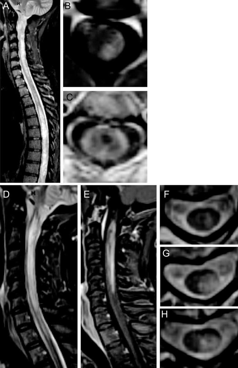
Figure Neuroimaging of CSF antibody-positive neuromyelitis optica
Case 1: Sagittal T2-weighted STIR MRI (A) shows hyperintensity throughout the cervical and thoracic spinal cord. Axial T1-weighted postgadolinium MRI at the level C2 (B) shows dorsal enhancement and at level C2–3 (C) shows peripheral enhancement and a central T1-weighted hypointensity. Case 2: Sagittal T2-weighted STIR MRI (D) shows hyperintensity from the lower medulla caudally with enhancement on T1-weighted postgadolinium MRI (E). Case 3: Axial T2-weighted MRI at successive levels C2 (F), C3 (G), and C4 (H) show central gray matter involvement.
