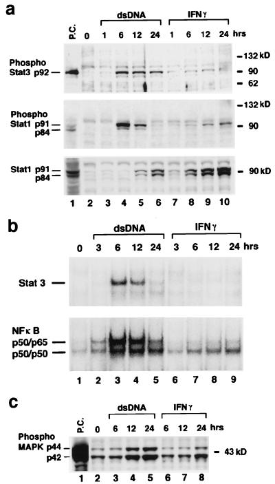
Figure 4.
dsDNA activates Stat1, Stat3, MAPK, and NF-κB. dsDNA transfection and IFN treatment of FRTL-5 cells were performed exactly as described for Figs. 1–3 by using 5 μg of poly(dI-dC)⋅poly(dI-dC). (a) Total cell lysate was prepared, and Western blot analysis was performed as described (20). Lane 1 (P.C.) contains a positive control cell lysate (New England Biolabs). (b) Nuclear protein was prepared, and gel-shift analysis was performed as described (15, 16, 19). Consensus ODNs for Stat3 and NF-κB are from Santa Cruz Biotechnology. (c) Western blot analysis with an antibody against phosphorylation-specific p44/p42 MAPK. Shown are typical results from at least four different experiments performed on different batches of cells.

