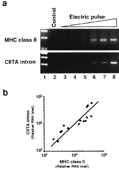
Figure 5.
Tissue damage by electric pulsing coordinately increases MHC gene expression and genomic DNA in the cytoplasm. FRTL-5 cells (5 × 10⁶ cells in Dulbecco’s PBS) were pulsed once with a Gene Pulser (Bio-Rad) set at 0.3 kV and at capacitances of 0.25, 25, 125, 250, and 960 μF or pulsed twice with a capacitance of 960 μF (lanes 3–8, respectively). Cells were washed with medium, returned to a 10-cm dish, and cultured for 48 h until RNA was recovered. Damage was estimated microscopically by trypan-blue exclusion and plating efficiency after pulsing. After two pulses at 960 μF, 60% of cells were fused or died. (a) Reverse transcription–PCR data compare MHC class II expression with contamination of total RNA preparations by leaked genomic DNA, measured by using PCR primers that detect an intron sequence of rat CIITA genome DNA (M. Pietrarelli, K.S., and L.D.K., unpublished results). Data are typical results from four different experiments performed on different batches of cells. (b) The correlation of MHC class II and CIITA intron levels for pulses eliciting significant increases of each (as shown in a, lanes 5–8) is presented after densitometry of the results. Data are mean values from four experiments.

