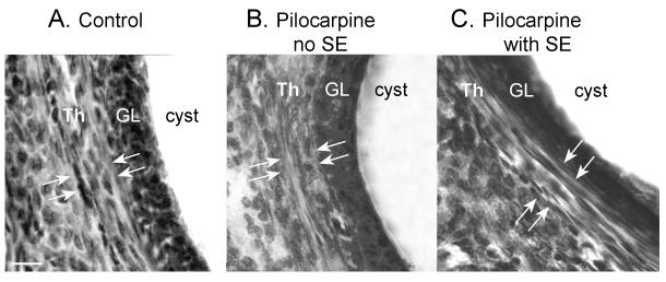
Figure 4. Lack of thecal cell hyperplasia in ovarian cysts of pilocarpine-treated rats.
Cystic follicles from a saline control (A), pilocarpine control (B), and epileptic rat (C). Hyperplasia of the thecal cell layer (Th) surrounding the granulosa cell layer (GL) was not detectable. Arrows demarcate the borders of the thecal cell layer. Calibration = 50 μm.

