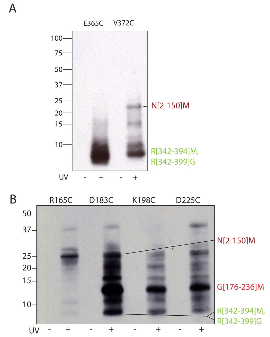
Figure 5. Interdomain crosslinking from σ4 and σ2.
Crosslinker was attached at the indicated residues within σ4 (A) and σ2 (B) through a disulfide. Irradiated (UV+) and non-irradiated (UV−) samples were digested with CNBr, separated by reducing SDS-PAGE (10–20% acrylamide, tris-tricine buffer system) and probed for biotin by Western blotting (HRP-streptavidin). The identity of the labeled bands is shown at right. The N[2–150]M, R[342–394]M and R[342–399]G bands were identified by mass spectrometry, the G[176–236]M band was identified as described in the text.

