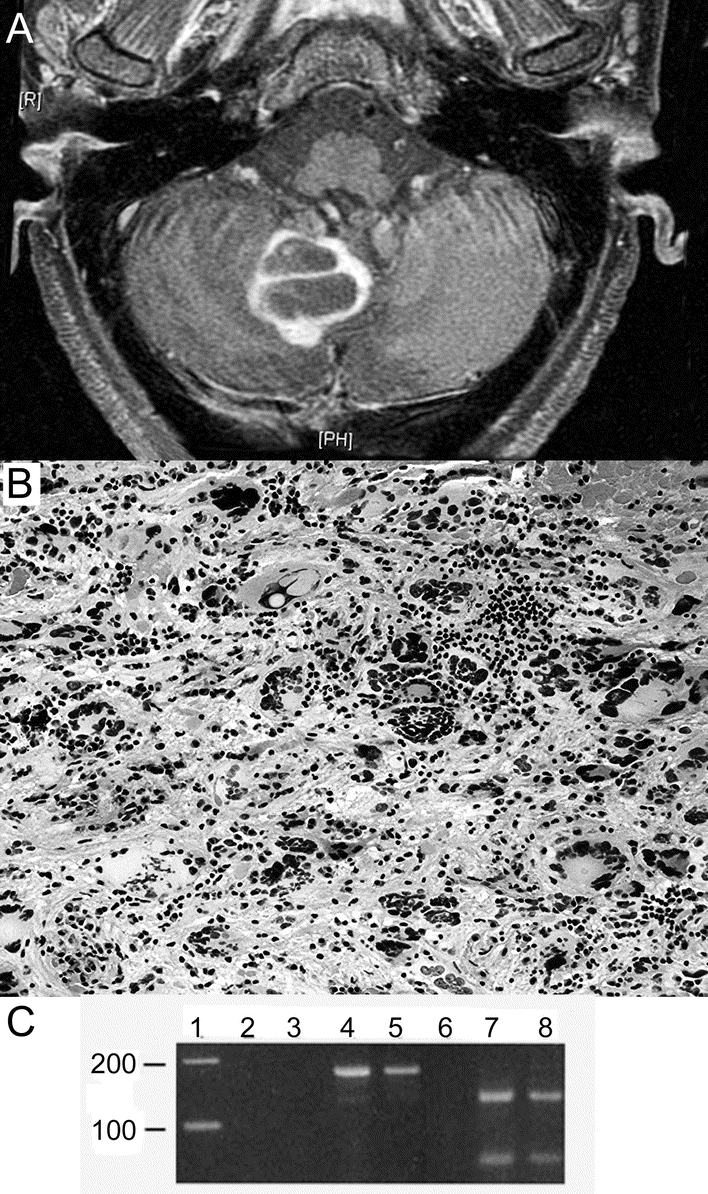
Figure MRI and histologic examination of the cerebellum
(A) Gadolinium-enhanced T1 FLAIR MR image demonstrating a large cystic ring-enhancing lesion in the context of high signal intensity lesions of the cerebellum. (B) Excisional biopsy of the cerebellum demonstrating bizarre multinucleated giant cells surrounded by an inflammatory cell infiltrate (hematoxylin-eosin, original magnification 100×). (C) Ethidium-bromide stained gel demonstrating presence of JCV in the patient’s pseudotumor. Formalin-fixed, paraffin-embedded sections of the patient’s lesion (lanes 4 and 7) and autopsy-derived progressive multifocal leukoencephalopathy (PML) (lanes 5 and 8) and normal brain (lanes 3 and 6) were used to extract DNA for PCR. Lanes 2, 3, 4, and 5 show the results of PCR amplification of a 173 base pair segment of polyoma virus in a reaction run without template DNA (lane 2), with normal brain DNA (lane 3), DNA from the patient’s lesion (lane 4), and from an unrelated case of PML (lane 5). Lanes 6, 7, and 8 display BamHI digests of the PCR products run in lanes 3, 4, and 5, respectively. Both the patient’s lesion and the case of PML show specific, 120 and 53 base pair fragments that occur only with JCV, which has a BamHI restriction site in the amplicon (SV40 and BKV do not share this restriction site). Lane 1 contains a 100 bp DNA ladder.
