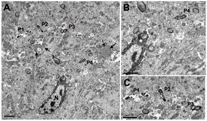
Fig. 6.
Electron micrographs of an Iba1-labeled microglial cell with Iba1-labeled processes in the hilus of the dentate gyrus. A shows an Iba1-labeled cell body with four cross-sectioned Iba1-labeled processes found approximately 4.6 μm (P4) and 11 μm (P1-P3) away from the cell’s nucleus (N). Mossy fibers (arrows) can also be seen scattered throughout the neuropil. B is an enlargement of the Iba1-labeled cell found in A and shows an inclusion body (asterisk) located its perikaryon. One of the Iba1-labeled processes (P4) is also shown. C is an enlargement of the three cross-sectioned Iba1-labeled processes (P1–P3). Note the arrangement of the cross-sectioned processes, demonstrating what appears to be a central process (P2) with two side branches (P1, P3). Scale bars = 2 μm for A–C.

