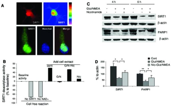
Fig. 3.
Nicotinamide treatment preserves levels of cellular NAD+ and SIRT1 in neurons subjected to an excitotoxic insult. a SIRT1 immunoreactivity in cultured cortical (upper panel) and hippocampal (lower panel) neurons. SIRT1 immunoreactivity was concentrated in the nucleus and so colocalized with Hoechst (DNA-binding dye) fluorescence. b SIRT1 deacetylase activities detected in different cell-free reactions or in nuclear proteins extracted from cortical cells that had been subjected to the indicated treatments. G/N glutamate and NMDA, Nic nicotinamide (2 mM). c Immunoblot showing SIRT1 and PARP-1 (full length PARP-1 band is shown) levels in cortical neurons treated with glutamate and NMDA alone or in combination with nicotinamide for 4 or 6 h. Blots were probed with β-actin. d Densitometric analysis (lower) of full length of PARP-1 and SIRT1 levels (normalized to the β-actin level) in cortical cells that had been exposed to glutamate/NMDA for 6 h. Values are the mean and SD of determinations from 4-6 cultures. *P < 0.05, **P < 0.01

