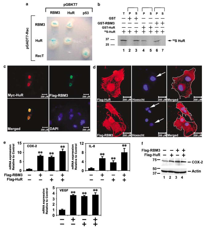
Figure 5.
RBM3 and HuR interact and enhance stability. (a) Yeast two-hybrid interaction of RBM3 with HuR. RBM3 and HuR expressed as bait and test proteins interact in the yeast by the colonies formed on quadruple dropout media. Breakdown of the X-α-gal results in a blue colony. Tumor suppressor protein p53 and SV40 T antigen (RecT) were used as positive control for interaction, but negative for interaction with either RBM3 or HuR. (b) GST pull-down assay. 35S-methionine labeled in vitro translated HuR (35S-HuR) was incubated with either GST-RBM3 or GST-HuR. The GST-proteins were immobilized on to glutathione sepharose beads. The immobilized proteins were separated by SDS–PAGE and subjected to phosphorimager analyses. Pure GST served as negative control. (c). Colocalization of HuR and RBM3. HeLa cells were transiently transfected with plasmids expressing myc-epitope tagged HuR and FLAG-epitope tagged RBM3. Immunocytochemistry was performed for the myc and FLAG epitopes. Images for the HuR and RBM3 were merged demonstrating colocalization. Nucleus was stained by DAPI. (d) Nuclear-cytoplasmic shuttling of HuR and RBM3. Plasmids encoding FLAG-epitope tagged HuR or RBM3 were transiently transfected into human HeLa cells and subsequently fused with mouse NIH3T3 cells. The proteins were immunostained for the FLAG tag, and the nuclei by Hoescht stain to differentiate human and mouse nuclei. Mouse nuclei, seen as punctuate staining are denoted by an arrow. (e) RBM3 and HuR induce COX-2, IL-8 and VEGF mRNA expression. Ectopic expression of Flag epitope-tagged RBM3 and HuR resulted in significant increase in endogenous COX-2, IL-8 and VEGF mRNA in HCT116 cells. There was a trend for even higher levels when proteins were coexpressed (**P<0.01). (f) COX-2 protein increased in cells expressing RBM3 and HuR. COX, cyclooxygenase; GST, glutathione S-transferase; P, GST-bound fraction; S, supernatant; SDS–PAGE, sodium dodecyl sulfate–polyacrylamide gel electrophoresis; T, total input or whole cell extract; VEGF, vascular endothelial growth factor.

