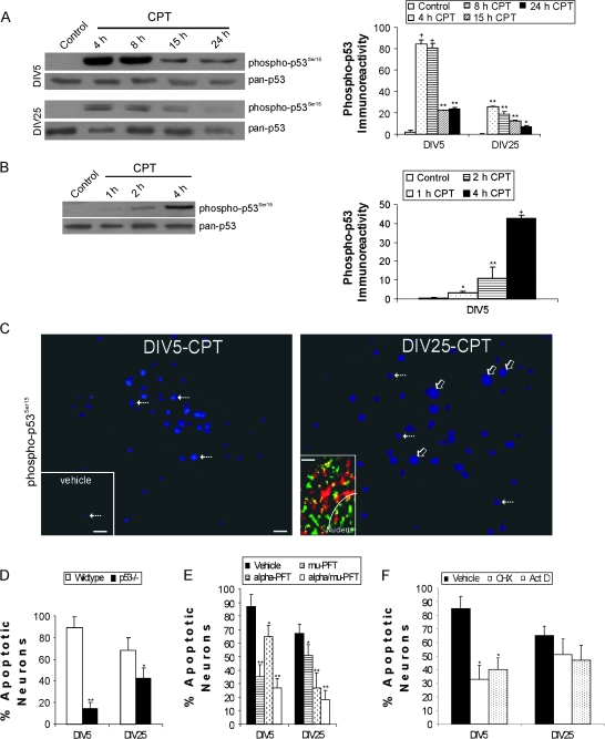
Figure 2.
p53 regulates DNA damage-induced apoptosis in immature and mature cortical neurons through partly different mechanisms. (A) Western blots showing robust activation of p53 through phosphorylation of serine-15 (detected with phosphorylation state-specific antibody) with 10 μM CPT treatment for 4 through 24 h (h) in DIV5 and DIV25 neurons (10 μg total protein loaded per lane). Total levels of p53 (detected with a phosphorylation state-independent pan antibody) remain stable or decrease over time. Graph shows the optical density measurements of phospho-p53 (in relative OD units) in DIV5 (immature) and DIV25 (mature-differentiated) neurons with 10 μM CPT treatment. Values are mean ± SD (n = 3 different cultures of 6 pooled 35 mm wells per data point). Symbols denote: significantly greater +P < 0.0001, **P < 0.001, and **P < 0.01 versus vehicle. (B) Western blot showing rapid activation of p53 by phosphorylation of serine-15 (detected with phosphorylation state-specific antibody) with 10 μM CPT treatment for 1 through 4 h in DIV5 neurons (7 μg total protein loaded per lane). Total levels of p53 (detected with a phosphorylation state-independent pan antibody) remain stable over this time course. Graph shows the optical density measurements of phospho-p53 (in relative OD units) in DIV5 neurons with 10 μM CPT treatment. Values are mean ± SD (n = 3 different cultures of 6 pooled 35 mm wells per data point). Symbols denote: significantly greater +P < 0.001, **P < 0.01, and *P < 0.05 versus vehicle. (C) Phospho-p53 immunoreactivity (cascade blue labeling) is present robustly at 4 h of CPT treatment in many DIV5 and DIV25 cortical neurons. In DIV5 neurons treated with CPT, phospho-p53 immunoreactivity is confined mostly to the nucleus (hatched arrows) in positive cells. In DIV25 neurons treated with CPT, phospho-p53 immunoreactivity is present throughout the cell body (nucleus and cytoplasm) in many neurons (open arrows), but in some neurons in the same microscopic field p53 immunoreactivity is confined to the nucleus (hatched arrows). Note that nuclei with p53 staining (hatched arrows) in DIV5 and DIV25 neurons have similar sizes, whereas the neurons with both cytoplasmic and nuclear labeling in DIV25 cultures are larger because the entire cell body is labeled. Scale bar in DIV5-CPT image = 10 μm (same bar for DIV-25 image). Only a few nuclei are positive for phospho-p53 (blue labeling) in vehicle control cultures (lower left inset, hatched arrow). Vehicle inset scale bar = 80 μm. Immunocytochemical dual labeling (lower left inset in right panel) for phospho-p53 (green) and the mitochondrial marker manganese superoxide dismutase (red) reveals a mitochondrial localization for some p53 (seen as yellow) in CPT-treated DIV25 neurons. The white curved line delineates partially the border between the nucleus and the cytoplasm (inset scale bar = 0.5 μm). (D) p53 gene deletion reduces apoptosis in DIV5 and DIV25 cortical neurons treated with CPT for 24 h. Values are mean ± SD based on at least 6 images of Hoechst-stained neurons per group and were replicated in 3 different culture experiments. Symbols denote: significantly lower **P < 0.001 and *P < 0.01 versus wild-type neurons. (E) Pharmacological inactivation of p53 reduces apoptosis in immature and mature wild-type cortical neurons. Neurons were treated (10 μM) with cyclic-α-pifithrin (α-PFT) that blocks reversibly p53-dependent transactivation of p53-dependent responsive genes, μ-PFT that blocks p53 interaction with Bcl-xL and Bcl-2 and inhibits selectively p53 translocation to mitochondria without affecting the transactivation function of p53, or a combination of both drugs for 2 h before exposure to 10 μM CPT. At 24 h, neurons were assessed for apoptosis. Values are mean ± SD based on at least 6 images of Hoechst-stained neurons per group and were replicated in 3 different culture experiments. Symbols denote: significantly lower **P < 0.001 and *P < 0.05 versus vehicle-treated neurons. (F) CPT-induced apoptosis of immature cortical neurons (but not mature cortical neurons) is transcriptionally and translationally dependent. Neurons were treated with the protein synthesis inhibitor cycloheximide (1 μg/ml) or the RNA synthesis inhibitor actinomycin D (1 μg/ml) for 2 h before exposure to 10 μM CPT. At 24 h, neurons were assessed for apoptosis. Values are mean ± SD based on at least 6 images of Hoechst-stained neurons per group and were replicated in 2 different culture experiments. Symbol denotes significantly lower *P < 0.001 versus vehicle-treated neurons.

