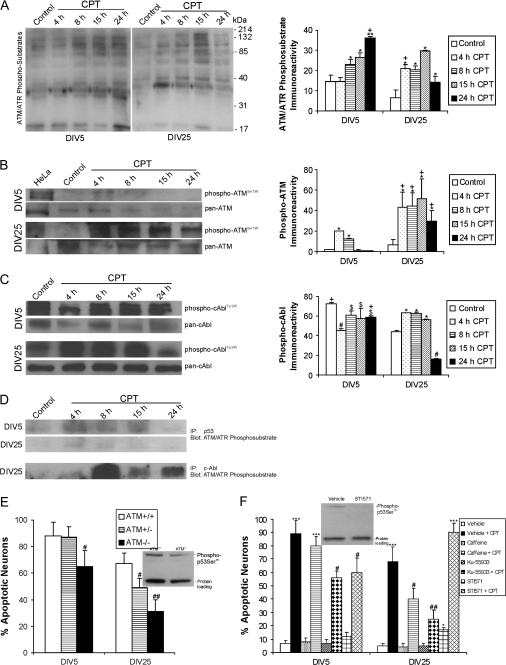
Figure 4.
DNA damage response kinases drive CPT-induced p53-mediated apoptosis in immature and mature cortical neurons. A. Western blots show activation of ATM/ATR kinase (by detection of phosphorylated substrate proteins) after CPT treatment (10 μM) for 4 through 24 h (h) in DIV5 and DIV25 neurons. Graph of ATM/ATR phosphosubstrate immunoreactivity (in relative OD units) in DIV5 (immature) and DIV25 (mature-differentiated) neurons with 10 μM CPT treatment. Values are mean ± SD (n = 3 different cultures of 6 pooled 35 mm wells per data point). Symbols denote: significantly greater **P < 0.001 and **P < 0.01 versus age-matched vehicle; significantly greater +P < 0.01 versus time-matched CPT-treated neurons. (B) Western blots show robust activation of ATM through phosphorylation of serine-1981 (detected with phosphorylation state-specific antibody) after 10 μM CPT treatment for 4 through 24 h in DIV5 and DIV25 neurons. Total levels of ATM (detected with a phosphorylation state-independent pan antibody) decrease over time. γ-Irradiated HeLa cells are a positive control for DNA damaged-induced activation of ATM. Graph of phospho-ATM levels (in relative OD units) in DIV5 (immature) and DIV25 (mature-differentiated) neurons with 10 μM CPT treatment. Values are mean ± SD (n = 3 different cultures of 6 pooled 35 mm wells per data point). Symbols denote: significantly greater *P < 0.01 versus age-matched vehicle; significantly greater +P < 0.05 than time-matched DIV5 neurons. (C) Western blots for activated tyrosine-245-phosphorylated c-Abl (detected with phosphorylation state-specific antibody) and total c-Abl (detected with a phosphorylation state-independent pan antibody) with 10 μM CPT treatment for 4 through 24 h (h) in DIV5 and DIV25 neurons. Graph of phospho-cAbl levels (in relative OD units) in DIV5 (immature) and DIV25 (mature-differentiated) neurons with 10 μM CPT treatment. Values are mean ± SD (n = 3 different cultures of 6 pooled 35 mm wells per data point). Symbols denote: significantly lower #P < 0.001 and $P < 0.05 versus age-matched vehicle-treated neurons; significantly greater *P < 0.01 versus age-matched vehicle-treated neurons; significantly greater +P < 0.01 than time-matched DIV25 neurons. (D) Immunoprecipitation assay identifying p53 and c-Abl as direct ATM/ATR targets in cortical neurons undergoing apoptosis induced by CPT. Results were confirmed in 3 different experiments. (E) ATM gene deletion provides significant protection against apoptosis in DIV5 and DIV25 cortical neurons treated with CPT for 24 h. Values are mean ± SD based on at least 6 images of Hoechst-stained neurons per group and were replicated in 3 different culture experiments. Symbols denote: significantly lower #P < 0.05 and ##P < 0.01 versus wild-type neurons. Western blot shows that ATM gene deletion attenuates p53 activation. (F) Pharmacological inactivation of ATM and c-Abl protects against apoptosis in immature and mature wild-type cortical neurons. Neurons were treated with highly specific (Ku-55933, 10 μM) or less specific (caffeine, 100 μM) ATM inhibitors or the c-Abl inhibitor (STI571, 10 μM) for 2 h before exposure to 10 μM CPT. At 24 h, the neurons were assessed for apoptosis. Values are mean ± SD derived from at least 6 images of Hoechst-stained neurons per group and were replicated in 3 different culture experiments. Symbols denote: significantly higher ***P < 0.001 and *P < 0.05 versus age-matched vehicle-treated neurons; significantly lower #P < 0.01 and ##P < 0.001 versus age-matched CPT/vehicle-treated neurons. Western blotting shows that p53 activation is attenuated by c-Abl inhibition. ATM inhibition also attenuated p53 activation (data not shown).

