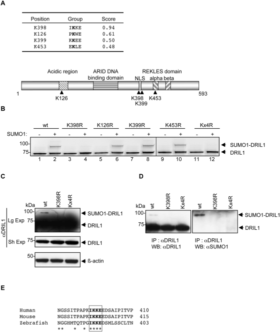
Figure 1. DRIL1 is sumoylated in vitro and in vivo.
(A) Table showing DRIL1 potential SUMO consensus motifs and SUMOplot™ score (Abgent; www.abgent.com/doc/sumoplot). Below is the schematic structure of DRIL1 showing lysines 126, 398, 399, 453 and the functional domains. (B) In vitro sumoylation assay performed with 35S-labeled in vitro-translated wt, SUMO point mutants (K126R, K398R, K399R, K453R) or the quadruple mutant (Kx4R) of DRIL1, incubated in a sumoylation mix containing purified E1, E2 and ATP in the absence or presence of SUMO1. (C) 293T cells transfected with wt DRIL1, K398R or Kx4R mutants. Lysates were Western blotted using antibodies against DRIL1 and β-actin as loading control. (Sh Exp) for short exposure and (Lg Exp) for long exposure. (D) 293T cells transfected with wt DRIL1, K398R or Kx4R. Lysate were immunoprecipited (IP) with antibody against DRIL1, and precipited proteins were western blotted (WB) using antibodies against DRIL1 (left panel) or SUMO1 (right panel). (E) Alignment of amino acid sequences of DRIL1 from human, mice and zebrafish spanning the conserved SUMO consensus motif using Clustal software.

