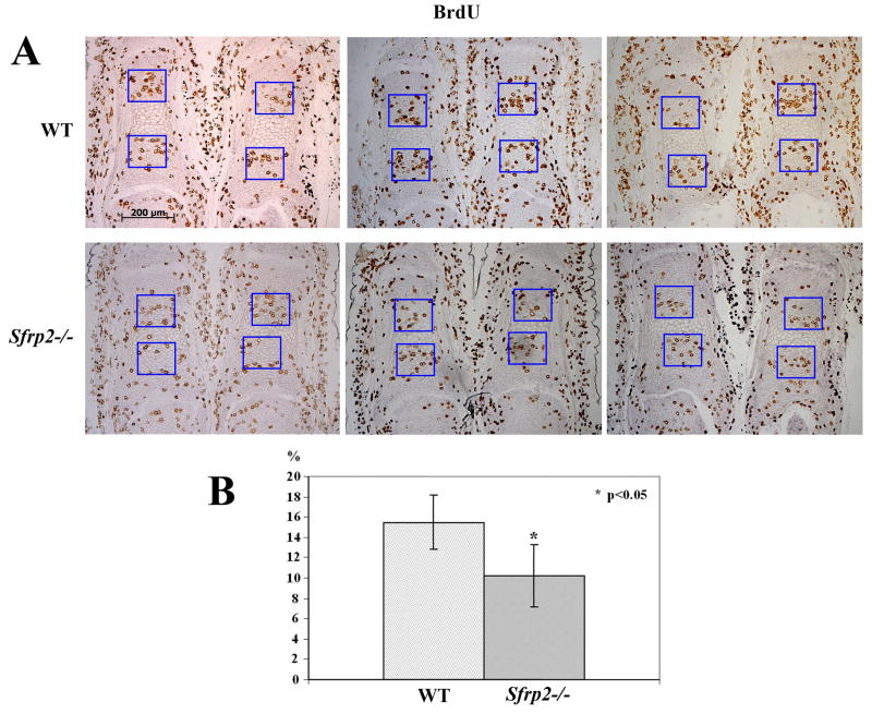
Figure 6.
Chondrocyte proliferation defect in Sfrp2-/- distal limb elements. (A) BrdU labeling shows decreased number of BrdU positive proliferating chondrocytes in E17.5 first phalanx sections of Sfrp2-/- versus controls. Also note the consistent hypertrophy delay in mutant phalanges. (B) Percentage of chondrocytes positively labeled with BrdU staining. The difference between WT and Sfrp2-/- mice (n=3) was statistically significant (p<0.05).

