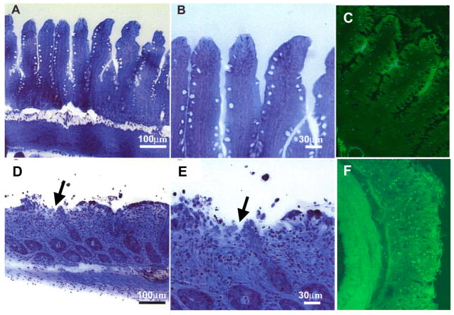
Figure 1. Auto-digestion of the intestine by pancreatic enzymes during ischemia.
Micrographs of rat intestinal wall morphology (semi-thin section stained with toluidine blue) and zymographic image with trypsin fluorescently quenched substrate (green fluorescence) before (Panels A, B, C, respectively) and after 45 min intestinal ischemia (D, E, F). Note the extensive damage to the microvilli and mucosal epithelium (D, E, arrows) and penetration of activated trypsin across the full thickness of the intestinal wall (F) with activation of trypsin activity (bright green fluorescence). Adapted from (29, 92).

