
Figure 4. Enhanced MMP activity in the SHR Microcirculation.
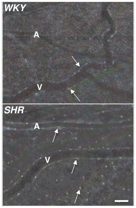
Digital fluorescent micrographs of WKY and SHR mesenteric microvessels labeled with fluorogenic peptide substrate showing matrix metalloproteinase (MMP-2, 9) enzymatic activity. Arterioles (A) and venules (V) are visible. Note the enhanced fluorescent emission over the endothelial cells and mast cells in the SHR, an affect that is less detectable after the doxycycline treatment. Adapted from (25). Bar equals 60 μm.