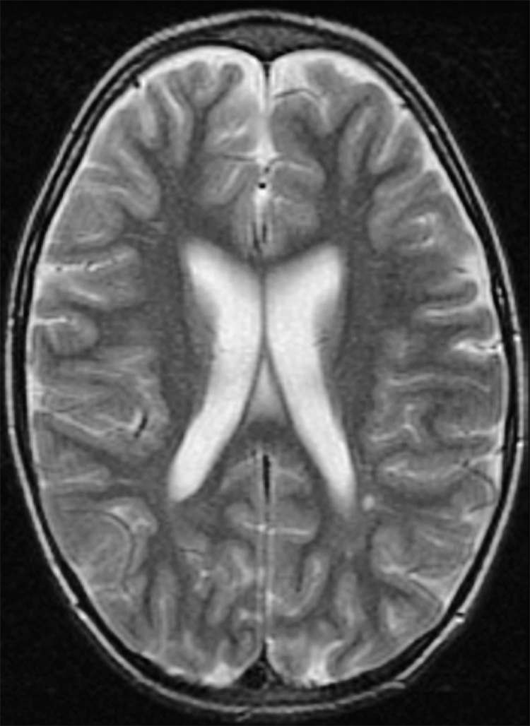
Figure 4.
Subject #204: Ventricular enlargement with volume loss and enlarged extra-axial spaces. Slice at level of ventricular measurement. Transverse T2-weighted fast spin echo MR image (TR/TE = 3000/102 ms, ETL = 12, slice thickness 5mm with 0mm gap interleaved, matrix = 256 × 192, FOV = 22 × 16cm).

