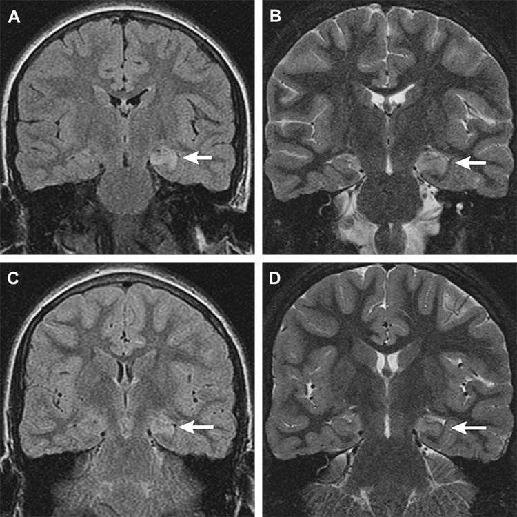
Figure 5.
Subject #209: Left hippocampal signal abnormality (arrows) in a ten-year-old. Coronal MR images, initial scan (a, b), and follow up 36 days later (c,d). (a) Increased signal left hippocampus, coronal fast fluid attenuated inversion recovery (FLAIR) MR image (TR/TE/TI = 10000/127/2250, slice thickness 4mm with 0mm gap interleaved, matrix = 256 × 192, FOV = 18cm). (b) Swelling and increased signal left hippocampus, coronal oblique fast multiplanar inversion recovery (FMPIR) MR image (TR/TE/TI = 5000/104/120ms, ETL =16, slice thickness/gap = 3mm/0mm, matrix = 512 × 256, FOV = 16cm). (c) Persistent increased signal left hippocampus, coronal FLAIR MR image (TR/TE/TI = 10000/142/2200, slice thickness 4mm with 0mm gap interleaved, matrix = 256 × 192, FOV = 18cm). (d) Subtle volume loss with persistent T2 prolongation left hippocampus, coronal FMPIR MR image (TR/TE/TI = 5000/96/120ms, ETL =16, slice thickness/gap = 3mm/0mm, matrix = 512 × 256, FOV = 16cm).

