
Figure 1. Kinetic differences in the effects of starvation-induced multi-site phosphorylation of Bcl-2 on the disruption of Bcl-2/Beclin 1 and Bcl-2 / Bax complexes.
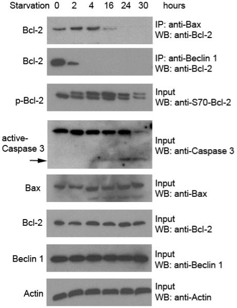
HeLa cells were starved in HBSS medium for the indicated time period. Bcl-2 co-immunoprecipitation with Bax (top panel) and Beclin 1 (second panel) was detected with an HRP-conjugated monoclonal anti-Bcl-2 (clone 100) antibody. Other panels represent the total amount of phospho-Bcl-2 (third panel), caspase 3 (fourth panel, arrow denotes active caspase 3, the lower molecular weight cleavage band), Bax (fifth panel), Bcl-2 (sixth panel), Beclin 1 (seventh panel) detected in cell lysates by Western blot analysis with the indicated antibody. Actin was used as a loading control (bottom panel).