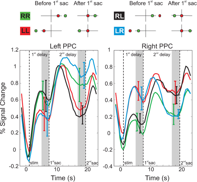
Figure 10.
Eye-centered reference frame revealed by fMRI. A subject fixates the origin while two targets are briefly flashed (before 1st saccade column). The final goal (green target) is flashed first, followed by the first fixation point (red target). The subject first makes a saccade to the red target (after 1st saccade column) and subsequently makes a saccade to the green target (not shown). In some conditions, the representation of the final goal (green target) is kept in the same hemisphere (e.g., goal stays in right hemisphere in the green RR condition and goal stays in the left hemisphere in the red LL condition). In other conditions, the saccade to the first fixation point (red target) causes the final goal (green target) to switch its location from one hemisphere to the other (e.g., from right to left in black RL condition and from left to right in blue LR condition). B. Activity that stays in the right hemisphere is shown by the green trace (RR condition), while activity in the left hemisphere is shown by the red trace (LL condition). Activity can be seen jumping from one hemisphere to the other with the black (RL) and blue (LR) conditions. For example, in the left PPC, the black trace follows the green trace during 1st delay period, but follow the red trace during the 2nd delay period. Replotted with permission from Medendorp et al., 2003a.

