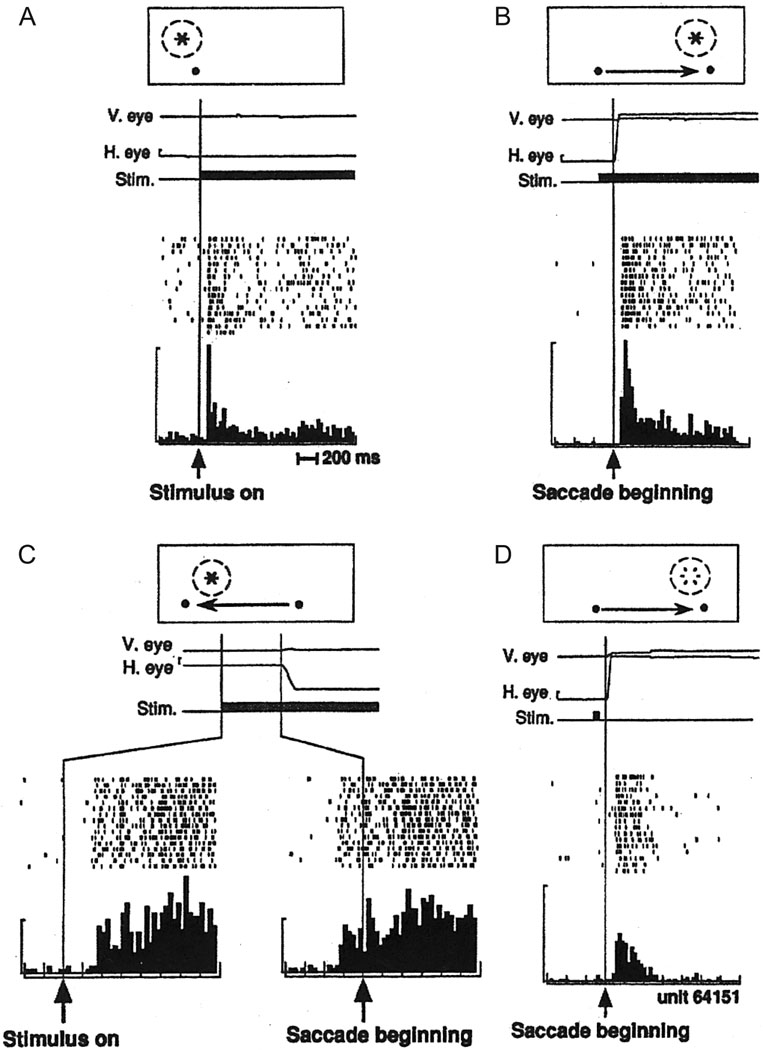
Figure 3.
Neural evidence for spatial updating in LIP. A. A cell in LIP responds when a visual target falls within its receptive field. Data aligned on visual stimulus onset. B. A cell in LIP responds when a saccadic eye movement brings the cell’s receptive field onto an illuminated target. Data aligned on saccade onset. C. Some cells begin to respond to an impending shift in the cell’s receptive field even before the eye movement begins. Bottom-right raster plot and histogram are aligned on the onset of the saccade. D. Some cells respond when their receptive field shifts to a location in which a visual target was previously illuminated. Open flash symbol indicates that the stimulus was extinguished before the eyes moved. Data aligned on saccade onset. Replotted with permission from Duhamel et al., 1992a.

