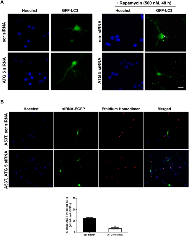
Figure 7. ATG 5 down-regulation decreases human A53T ASYN-induced death in rat cortical neurons.
(A) Five day-old cortical cultures were transfected with GFP-LC3 cDNA together with the scrambled (scr) or the ATG 5 siRNA. 24 hrs later, rapamycin was added (500 nM, 48 hrs) and formation of GFP-LC3 vacuoles (dots) was determined by fluorescent microscopy. Representative fluorescent microscopic pictures showing the formation of GFP-LC3 vacuoles (indicated by the arrow) in cultures transfected with the GFP-LC3 construct together with the scr siRNA after rapamycin addition are shown. Vacuoles (dots) failed to be observed in rapamycin-treated cultures transfected with the GFP-LC3 construct together with the ATG 5 siRNA. (B) Five day-old cortical cultures were transduced with A53T ASYN adenovirus and 24 hrs later were transiently transfected with the scr or ATG siRNA along with an EGFP construct to monitor transfection. Cell death was assessed 72 hrs later by counting the percentage of EGFP-positive transfected cells that were positive for Ethidium Homodimer stain (dying cells). Representative pictures are shown in the upper panel and quantification of the percentage of scr or ATG5/EGFP positive cells that were also stained with Ethidium Homodimer is shown in the bottom panel. At least 100 EGFP-positive cells were counted per condition. The data are presented as mean±SE of 3 independent experiments (**p<0.01, Student's t-test, comparing A53T ASYN transduced neurons transfected with the scr or the ATG 5 siRNA. Bars: (A) 10 µM; (B) 50 µM.

