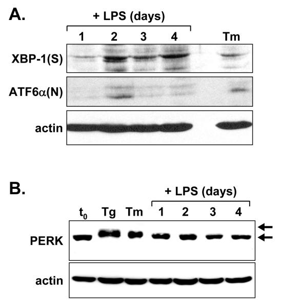
Figure 2.
Immunoblot analysis of UPR components in LPS-stimulated splenic B-cells. Splenic B-cells were cultured in the presence of LPS for the indicated intervals. At day 3, a portion of the cells was treated either for 5 h with 1 μg/ml tunicamycin (Tm) or 1 h with 0.4 μM thapsigargin (Tg) to provide positive controls for UPR activation. Cell lysates were prepared and equal cell equivalents were assessed by immunoblotting for A, XBP-1(S) and ATF6α(N) and B, PERK. Phosphorylated and nonphosphorylated forms of PERK are denoted by upper and lower arrows, respectively. Actin was assessed as a loading control.

