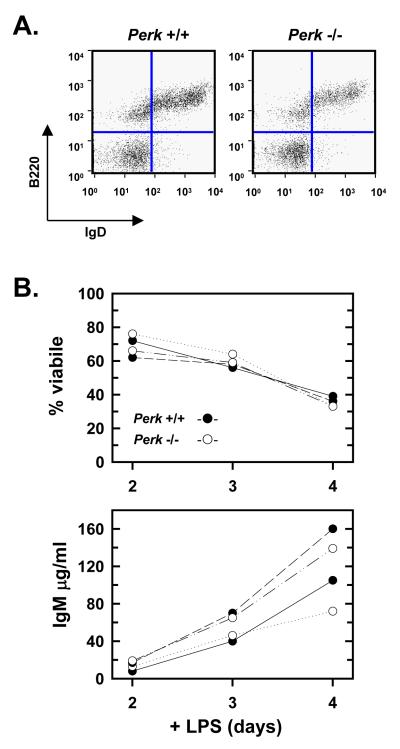
Figure 3.
Development and LPS response of PERK-deficient B-cells. Splenocytes and splenic B-cells were prepared from age- and sex-matched Perk+/+ and Perk-/- mice. A, Splenocytes were prepared, stained with FITC-conjugated anti-B220 and biotin-conjugated anti-IgD plus streptavidin-APC, and assessed by flow cytometry. Splenocytes scoring as B220+IgD+ mature B-cells appear in the upper right quadrants. Five mice for each genotype were analyzed and representative data are shown. B, Splenic B-cells from two separate Perk+/+ mice (●) and two separate Perk-/- mice (○) were cultured in the presence of LPS. At the indicated intervals, cell viability was assessed by trypan blue dye exclusion (upper panel) and the amounts of IgM present in the culture supernatants were determined by ELISA (lower panel).

