Figure 5.
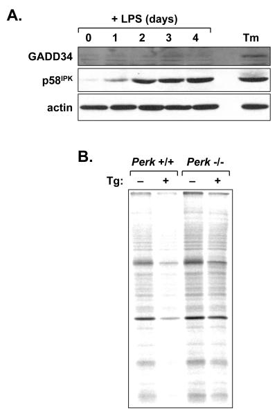
Induction of PERK regulators and responsiveness of the PERK pathway in LPS-stimulated B-cells. A, Splenic B-cells were cultured in the presence of LPS for the indicated intervals. At day 3, a portion of the cells was treated for 5 h with 1 μg/ml tunicamycin (Tm) to provide a positive control for UPR activation. Cell lysates were prepared and equal cell equivalents were assessed by immunoblotting for GADD34, p58IPK and actin as a loading control. B, Splenic B-cells were prepared from age- and sex-matched Perk+/+ and Perk-/- mice and cultured in the presence of LPS for 3 days. Cells were then treated for 1 h with vehicle alone or 1 μM thapsigargin (Tg) and metabolically labeled with 100μCi/ml [35S]methionine and cysteine for 10 min. Equivalent amounts of cell lysate (2×10⁵ cells) were resolved by SDS-PAGE under reducing conditions and signals from labeled proteins were captured by phosphorimaging. Thapsigargin treatment resulted in a 40% decrease in protein synthesis in +/+ cells and a 14% decrease in -/- cells.

