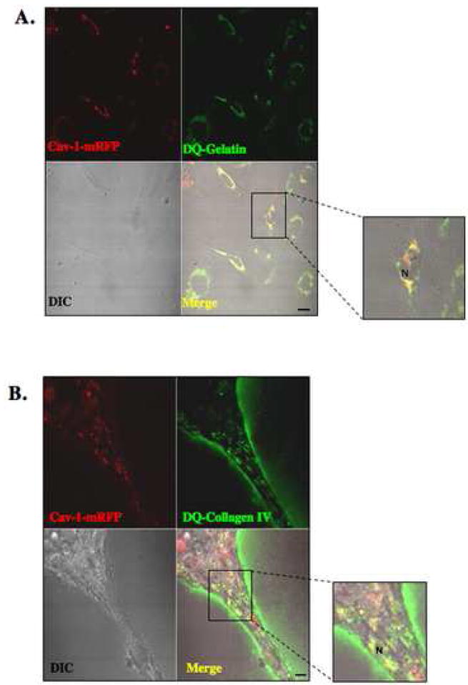
Figure 2. Intracellular colocalization of cav-1 with gelatin and collagen IV degradation products in HUVEC.
Equal numbers of HUVEC transfected with cav-1-mRFP construct were grown for 16 h on glass coverslips coated with either gelatin containing 25 μg/ml DQ-gelatin (A) or rBM (Cultrex) containing 25 μg/ml DQ-collagen IV (B). After 16 h, confocal images were taken of live cells (DIC), cav-1-mRFP (red) and DQ-substrate degradation products (green). DQ-substrate degradation products (green) are present intracellularly in HUVEC grown on gelatin and both intracellularly and pericellularly in HUVEC grown on rBM (see inserts). Colocalization of cav-1-mRFP and DQ-substrate degradation products appears yellow in the merged images. N, nucleus. Bar, 20 μm.

