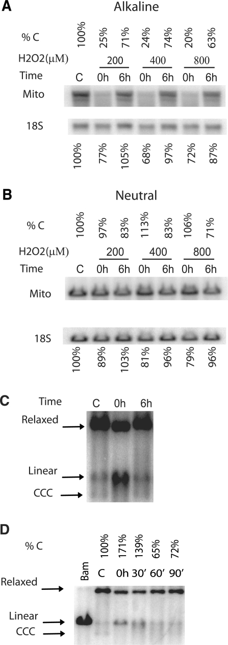
Figure 2.
Oxidative damage results in the degradation of mtDNA. (A and B) HCT116 cells were either left untreated (C, control) or treated with the indicated concentrations of H2O2 for 1 h, after which total genomic DNA was either extracted immediately (0 h), or allowed to repair for 6 h (6 h) prior to extraction. The DNA was digested with BamHI, which has a single recognition site in human mtDNA, quantitated and separated in 0.6% agarose gel under either alkaline (A) or neutral (B) conditions. After Southern blotting with mitochondrial (Mito) and nuclear (18S) DNA probes, the intensity of bands corresponding to intact mtDNA was expressed as a percent of control (untreated) values. (C) Oxidative DNA damage is accompanied by accumulation of linear mtDNA intermediates. HCT116 cells were treated with 200 µM H2O2, total DNA was extracted, digested with Bgl II, and subjected to Southern blotting under the neutral conditions. Bgl II has no recognition sites in mtDNA, and therefore the banding pattern is reflective of the native state of mtDNA. The positions of markers corresponding to covalently closed circular (CCC), linear and relaxed mtDNA molecules are indicated by arrows. (D) A time course of degradation of linear mtDNA intermediates. HCT116 cells were either left untreated (C) or treated with 400 μM H2O2 for 30 min, and then either lysed immediately, or allowed to recover for 30, 60, or 90 min. The percentage of linear mtDNA as compared to untreated control is indicated.

