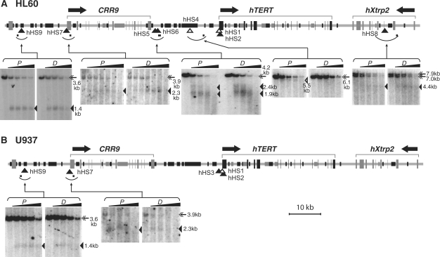
Figure 3.
DNase I hypersensitive sites at the hTERT locus and neighboring loci in HL60 cells (A) and U937 cells (B). HL60 and U937 cells were treated with DMSO for 4 days and TPA for 24 h, respectively. Nuclei were isolated and digested with 0, 2, 4, 8 and 16 U/ml DNase I in all panels except for the hHS7 panel of HL60 cells, in which 0, 0.5, 1, 2, 4, 8 and 16 U/ml DNase I were used. Genomic DNAs were extracted and digested with restriction enzymes, followed by Southern blotting and indirect labeling of genomic DNA bands. The top portion of each panel set is a schematic diagram of the genomic region containing the hTERT, hCRR9 and hXtrp2 loci. Horizontal arrows above the diagrams indicate the direction of transcription for each gene. Exons are depicted as tall rectangles and vertical lines. Black portions of intergenic and intronic sequences correspond to short repetitive sequences and dark gray portions represent mini-satellite sequences. Arcs below genomic sequences indicate chromosomal fragments that were examined by Southern analyses and small horizontal bars within the arcs denote the positions of probes for indirect labeling of the hypersensitive bands. In the Southern autoradiographs, proliferating cells (P) are shown on the left panels and differentiated cells (D) are on the right panels for each examined genomic region. Full-length genomic fragments are marked by double arrows. Open triangles point to DHS bands that appeared only in proliferating cells. The positions of these DHSs are also designated by open triangles in the genomic diagrams. Closed triangles indicate the hypersensitive bands and their genomic positions for constitutive DHSs that were present in both proliferating and differentiated cells. The double full-length genomic DNA bands in the hHS8 panel were caused by polymorphic mini-satellite sequences within the restriction fragment in HL60 cells. The sizes of full-length fragments and DHS bands were labeled on the right side of each panel set. Restriction digestions of genomic DNAs are as follows: hHS4, EcoRI; hHS5 and hHS6, EcoRI/SphI; hHS7, SacI; hHS8, SphI; and hHS9, EcoRI/SphI.

