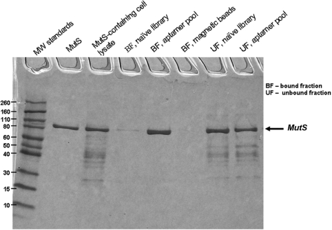
Figure 4.
SDS–PAGE with Coomassie staining of affinity pull-down of MutS from the E. coli cell lysate using the aptamers developed by AptaPIC. The lanes from left to right correspond to: (i) molecular weight standards (×1000); (ii) pure MutS; (iii) MutS-containing cell lysate, the sample contained 91.25 µg/ml and 18 µg/ml of MutS; (iv) MutS purification using the naive DNA library, bound fraction; (v) MutS purification using the developed aptamer pool, bound fraction; (vi) fraction bound to streptavidin-coated magnetic beads; (vii) MutS purification using the naive DNA library, unbound fraction; and (viii) MutS purification using the developed aptamer pool, unbound fraction.

