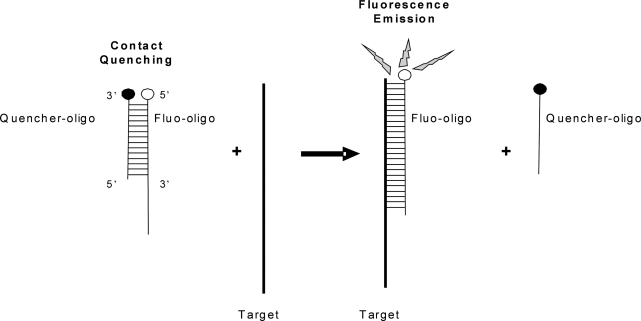
Figure 2.
Schematic representation of the broken beacon. The fluorescent oligonucleotide (fluo-oligo) forms a duplex with the quencher oligonucleotide (quencher-oligo); the proximity of the fluorophore and the quencher prevents fluorescence emission. In the presence of the target sequence, the quencher-oligo is displaced, allowing the fluo-oligo to emit.

