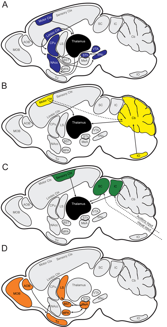
Fig. 12.
Schematic hypothesis for circuitry modulated by Foxp2 in the adult brain. A: Striatal modulation of fine motor response. B: Olivocerebellar modulation of motor timing. C: Thalamic integration of auditory and visual inputs. D: Limbic processing of olfactory input. Solid arrows indicate connectivity between regions in which Foxp2 expression is strong and continuous; broken arrows indicate pathways with discontinuous or weak expression. Connectivity between regions in which Foxp2 is not reciprocally expressed is not shown. AOB, accessory olfactory bulb; BST, bed nucleus of stria terminalis; Cb, cerebellum; CPu, caudate putamen; Ctx, cortex; IO, inferior olivary complex; LS, lateral septum; MeA, medial amygdala; MOB, main olfactory bulb; MPA, medial preoptic area; NAcc, nucleus accumbens; SNc, substantia nigra pars compacta; STh, subthalamic nucleus; Tu, olfactory tubercle; VTA, ventral tegmental area.

