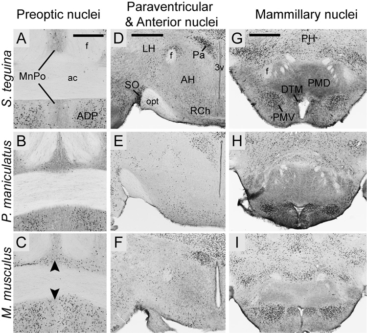
Fig. 7.
Foxp2 expression in selected hypothalamic nuclei in coronal sections from S. teguina, P. maniculatus and M. musculus. A–C: Expression in the midline median preoptic nucleus (MnPO) is pronounced in M. musculus (arrows in C) and minimal in P. maniculatus (B) and S. teguina (A). Expression in the anterodorsal nucleus (ADP) of the preoptic area is conserved across species. D–F: In all species, expression is concentrated in the paraventricular nuclei (Pa) and the supraoptic nucleus (SO), diffuse in the retrochiasmatic area (RCh) and absent from the anterior hypothalamic nuclei (AH). D–F: Foxp2 in mammillary nuclei, showing concentrated expression in dorsal tuberomammillary (DTM) and ventral premammillary (PMV) nuclei, and lack of expression in dorsal premammillary nucleus (PMD). Expression in S. teguina (A, D, G) is representative of S. xerampelinus. Scale bars = 300 µm in A (applies to A–C); 500 µm in D (applies to D–F); 500 µm in G (applies to G–I). f, fornix; ac, anterior commisure; 3V, third ventricle; opt, optic tract; PH, posterior hypothalamus.

