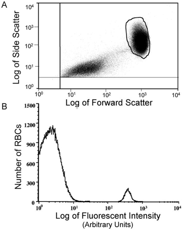
Figure 1.

Flow Cytometric Results for a Mixture of Unlabeled Red Blood Cells (RBCs) and Biotin-Labeled RBCs from a Representative Sample. Panel A: Enumeration of RBCs. The selected region (closed loop) includes unlabeled and labeled RBCs but excludes photomultiplier noise, platelets and any cellular fragments (left lower quadrant). Panel B: Histogram of unlabeled RBCs (large peak) and labeled RBCs (small peak) complexed with fluorescein-conjugated avidin reveals a complete separation between labeled and unlabeled RBCs allowing unambiguous enumeration.
