
Figure 1. Stimulus-induced Fos expression in the LC after SNI.
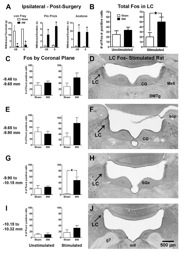
Panel A: Tactile allodynia (drop in von Frey threshold), mechanical hyperalgesia (increased response to pin prick), and cold allodynia (increased response to topical acetone) in rats with spared nerve injury (SNI) or sham surgery. There was no difference in hypersensitivity among SNI rats that were later assigned to the unstimulated (US) or stimulated with non-noxious stroking of the footpad (S) condition. Panel B: The total number of Fos-immunoreactive (Fos-ir) profiles in the LC (both sides) in sham and SNI rats 2 wk following SNI. Innocuous tactile footpad stimulation increased Fos in SNI rats. Panels C, E, G, I: The number of Fos-ir profiles in 4 distinct coronal divisions throughout the rostro-caudal extent of the LC. The effect of footpad stimulation in SNI rats reached statistical significance in the -9.90 to -10.15 mm division. Panels D, F, H, J: Representative photomicrographs of stimulated Fos-ir at each coronal division of the LC in an SNI rat. CG=central gray; DMTg=dorsomedial tegmental area; g7=facial nerve; Me5=mesencephalic 5th nucleus; mlf=medial longitudinal fasciculus; scp=superior cerebellar peduncle; SGe=supragenual nucleus. Magnification = 3X. Data is expressed as mean ± SEM. n=3-12. ★p < 0.05.