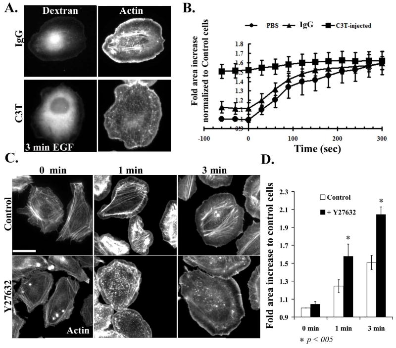
Figure 2. Inhibition of Rho/ROCK leads to an increase in cell area.
A) Starved cells were microinjected with 488 Alexa-Dextran along with IgG control (upper panels) or with 150 μg/ml C3T along with Dextran (lower panels), allowed to recover for 1h, then fixed and stained with Rhodamine phalloidin. Left-hand panels show the 488Alexa fluorescence in microinjected cells and right-hand panels show f-actin staining. B) Control, IgG/dextran or C3T/dextran injected cells were stimulated with EGF and time lapses were performed, collecting images every 20 seconds. The surface area of control or injected cells was determined, and normalized to cells at time 0 before stimulation. Data are the mean +/− SEM from 20 cells. C) MTLn3 cells, plated on collagen, were starved for 3 hours and treated without or with 25 μM of Y27632 for 30 min. Cells are then stimulated with EGF for 0, 1 and 3 min, fixed and stained with Rhodamine Phalloidin. D) Quantitation of the surface areas of cells treated as in panel C, plotted as fold increase relative to control cells. The data are the mean from 3 +/− SEM from 3 different experiments (15 cells/experiment). Scale bar is 10 μm.

