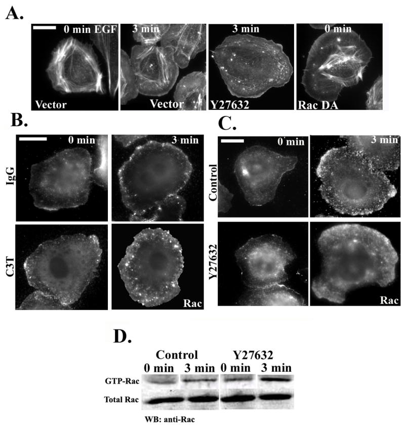
Figure 3. Inhibition of RhoA/ROCK leads to Rac hyperactivation and increased membrane localization.

A) Rhodamine phalloidin staining of cells transfected with vector alone (first two panels), transfected with vector and treated with Y27632 (third panel) or transfected with RacQ61L (last panel to the right). Cells were starved for 3 hours, stimulated with EGF for 0 or 3 minutes, and stained with Rhodamine phalloidin. B) Representative micrographs of starved cells microinjected with 488Alexa-Dextran and IgG (upper panels) or 150 μg/ml C3T (lower panels). Cells were stimulated with EGF for 0 or 3 minutes and stained with anti-Rac antibodies. C) Representative micrographs of starved cells treated without or with carrier (upper panels) or Y27632 (25 μM; lower panels) for 30 min. Cells were stimulated with EGF for 0 or 3 minutes and stained with anti-Rac antibodies. D) GST-CRIB pull-down from control or Y27632-treated cells were blotted with anti-Rac antibody. The lanes for each blot (GTP and total Rac) were taken from the same gel.
