
Figure 5. EGF-stimulated protrusion in Y27632-treated cells is Rac dependent.
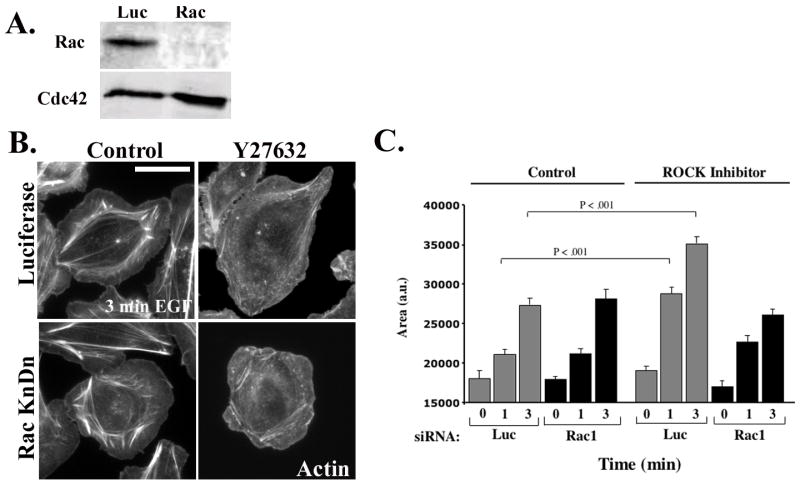
A) MTLn3 cells were transfected with control or Rac siRNA. Representative Western Blot showing the expression of Rac (upper panel) and Cdc42 (lower panel). B) Luciferase (upper panels) and Rac1 (lower panels) siRNA-treated cells were either incubated without or with 25 μM Y27632 for 30 min. The cells were then stimulated for 0, 1 and 3 minutes with EGF. The representative micrographs show cells stimulated for 3 minutes and stained with Rhodamine Phalloidin C) Quantitation of cells from (B) stimulated with EGF for 0,1 or 3 min. The data are normalized to the area of unstimulated control cells, and are the mean +/− SEM from at least 45 cells from three distinct experiments. Scale bar is 10 μm.