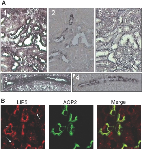
Figure 2.
Localization of LIP5 in mouse kidney. (A) Alternating mouse kidney sections were used to visualize the co-localization of LIP5 mRNA (by in situ hybridization; 1 and 3) and AQP2 (by immunohistochemistry; 2 and 4). LIP5 mRNA is detected in most epithelial cells. In situ hybridization using a sense probe did not reveal any specific staining (5). (B) Renal sections of mice receiving water ad libitum were subjected to immunohistochemistry for LIP5 (red) and AQP2 (green). In renal principal cells, which express AQP2, LIP5 shows similar localization to that of AQP2 (middle). In intercalating cells (*) and epithelial cells of other tubules (arrows), LIP5 staining is more punctuate.

